Portfolio
This portfolio is a collection of cases that have been planned, presented, and/or completed during my third and fourth years at The University of Maryland School of Dentistry.

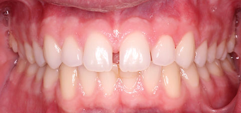

20 year old male presented with the chief complaint of "I don't like the size or the spaces in my front teeth." The patient was presented options and elected composite Veneers 7-10. The patient was not interested at this time to prep his teeth for veneers. A prep-less approach of composite veneers was done to fulfill the patient's chief complaint.


Student Dr. Morabito is thoroughly detailed and displays rational judgment regarding various cases. After a major dental operation, he extends personal follow-up care via phone calls. He is caring and will be an awesome dentist.
-Manny
Mrs. P presented to screening to with the chief complaint to have her lower anteriors fixed, however a thorough examination revealed an irritated palate. The differential diagnosis was denture-induced stomatitis and oral candidiasis. Nystatin Oral suspension [100,000units/ml] was prescribed in addition to Nystatin powder. The patient was instructed to use the oral suspension 2-3 times a day in addition to sprinkling the powder over the denture before placed back in her mouth. The patient was also instructed to soak the denture in a dilute bleach solution and to gently scrub the palate with a wash cloth daily. The Photos below are 2 week increments.


Photo at first visit
2 week follow-up
4 week follow-up
6 week follow-up
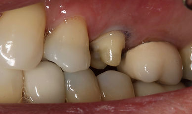
The patient had decay at the margin of #30 abutment tooth, it was repaired by removing the decay and placing amalgam and a final polish.

Decay on #30 margin

#30 preparation

#30 with amalgam placed

#30-Buccal amalgam polished
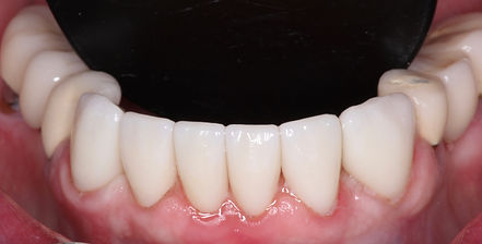
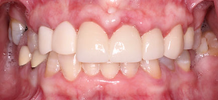
Mrs. P presented with wanting to have her lower teeth fixed to "look prettier." She presented with a maxillary denture and a bridge on the lower right and left. Diastema were also present between the mandibular canines and laterals and a prophylaxis was performed removing calculus from her lower anteriors revealing remaining tooth structure available to design her crowns.

To give a full smile showing both my upper and lower teeth is something I thought I would never experience. But, thanks to your hard work I am able to give that huge smile.
Words cannot express my gratitude. I look forward to having you as my dentist for life upon your graduation.
-Mrs. S.


Patient initial présentation
Patient presentation post prophylaxis
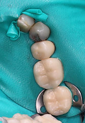
A gingivectomy was performed in preparation for the 6 crowns canine to canine on the lower to allow for for adequate ferrule. A wax up was fabricated to help the pt visualize the final outcome.


Patient mounted cast in occlusion
Mock up before preps

Prep guide with preps
Final crowns 22-27
Patient mounted cast slightly open

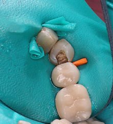
Gingivectomy was performed between 22-23 and 26-27 to help shape the future papilla.

Final preps
Final crowns 22-27

Patient presented with a temporary cantilever bridge 6-7. Treatment options were presented and patient elected to removed crown #8 and fabricate bridge from 6-8.


Patient presents with the chief complaint of wanting to fill the missing teeth. A treatment plan was accepted for Maxillary and Mandibular Cast Partials. Before initiation of the definitive treatment the disease control phase must be completed. The following pictures show lesions before, prepared, and finalized with composite restorations.
#26 Presented with a distal facial cavity/ secondary caries which appeared to be a defective restoration. The restoration was prepared and finalized with TPH composite.

Initial presentation of #26-DF

#26-DF preparation

#26 Final restoration
#9-DFL was a defective restoration was recurrent caries around the gingival margin.

Initial presentation #9 DFL

#9 cavity preparation

#9 Final restoration lingual view

#9 final restoration facial view
#28-DO was a defective restoration was recurrent caries around the distal margin
Initial presentation #28
#28 cavity preparation

#28 Final restoration

Patient presented for a 6 month recall. The patient had no chief complaint and had not desire for more treatment. Patient did however present with 2 defective restorations which were restored; #6 and #7 Class V.
Retracted smile

Initial presentation #6, #7

#6 preparation

#7 preparation

#6, #7 Final restoration

Initial presentation, fractured #8 and #9
Patient presented with the chief complaint of wanting to replace his front teeth. #8 and #9 are previously root canal treated and restored with crowns. It is suspected that the crowns fractured off due to the patient's deep bite and maligned lower anteriors. An Essix retainer with denture teeth was fabricated for esthetics as the patient completed disease control and ortho consult.
Patient presented with three defective restorations on the lower left.

Delivery of Essix retainer
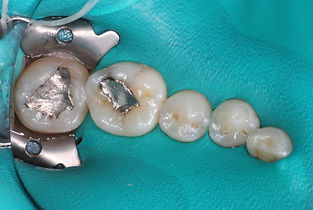
Initial presentation of 18-BO, 19-O, 20-O

Preparation of 18-BO, 19-O became a 19-BO, and 20-O
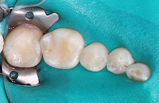
Final restorations of 18-BO, 19-BO, and 20-O
Patient presents with the chief complaint of "I want to have implants to hold my lower denture in, and I don't like these two teeth." The patient pointed to the chip on #9 and the discoloration of the class v on #10. #9 was repaired with composite and #11 was presented with recurrent decay which was repaired by removing and placing a new restoration.


Final restorations #9-FIL and #11- class v
Initial presentation of #9 and #11
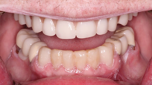
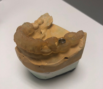
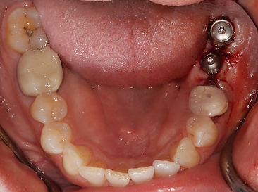
The patient's chief complaint of wanting implants to hold her lower denture was addressed with implants. It was planned for 4 implants initially however based on the DX imaging it would only be possible to place implants in the positions of 23 and 26. You will see the DX imagining with a denture duplicated radiographic guide, post-op radiographs and the final conversion of the lower denture to an implant-retained overdenture.

DX Pano pre-op

Existing lower partial was duplicated and radiographic guide fabricated using ortho resin and gutta-percha used as radiographic markers

Existing lower partial was duplicated

Radiographic guide scanned with the CBCT to aid in the implant placement plan

Post-op pano

Post-op of implants with locators.
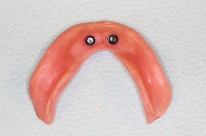
Post-op of lower denture retrofitted with housing

Full face smile at treatment planning appointment.

Clinical presentation of upper left, red arrow points to source of pain. Tooth 13 was rotated 90 degrees towards towards the posterior.

Patient wax-up of planned crowns.
Mrs. S presented to the clinic with the chief complaint of "I want to fix my front teeth." She was in an accident on a Friday and presented to the Dental school on Monday in Urgent care. An exam and radiographs revealed #8 class 4 crown fracture, #9 partially avulsed, and #10 presented with mobility class 1. Due to the nature of the avulsion of #9, it was extracted. The patient was worked up with multiple treatment plans with the main focus of esthetics repairing her smile. A wax-up was used to show the patient how her smile could be fixed.
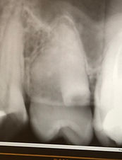
Premolar PA presentation of the "retained root"

The fused root on the distal root surface

Fused root on distal root surface
Mrs. S presented to the clinic with a second chief complaint 3 months after the disease control had begun of "Pain on the upper left." The pain was keeping her up at night. During the clinical examination and diagnostic test, it was revealed that the source of the pain is what appeared to be a supernumerary retained root. Upon elevation it was noticed that the adjacent tooth #13 move simultaneously with the retained root. With a closer look at the radiographic and clinical presentation it was predicted to be a fused root. The extraction of #13 did in fact reveal that the retained root was fused.


4 months after the trauma to the anterior teeth diagnostic testing was performed and revealed Necrotic pulp with asymptomatic periapical periodontitis. Radiograph revealed radiolucency around the apex. A root canal was completed.


The patient presented with Pain on the UL. The exam revealed a large carious lesion into the pulp. Dx Irreversible Pulpitis with asymptomatic periapical periodontitis.
Rct was performed with a build-up and an Emax Cerec designed crown


93 year old female presented with CC: "I want a new pair of dentures, I cannot chew food because the teeth are flat." New C/C dentures were made with most esthetic color of teeth and 10 degree posterior teeth to address the patient's chief complaint.


Upper immediate maxillary denture wax-up before processing

84 year old male presented with CC "I can't chew very well, i would like to replace my missing teeth. Treatment options were presented and the patient elected for Immediate Maxillary denture and immediate mandibular overdenture with 22 and 27 as abutment teeth. The immediate dentures were planned to be staged. It was also planned to fabricate Complete upper and lower dentures after 6 months of healing and bone remodeling.

Maxilla after extractions and alveoloplasty, tuberosities were not reduced in order to add retention with the loss of the right buccal plate

Upper immediate maxillary denture at 24-hour follow-up. The denture was relined and sore spots were relieved
65 year old female presents with the chief complaint "I would like to have implants to chew better."


26 year old female wisdom teeth removal. Lower wisdom teeth were partially boney impacted.


